Our Vitrectomy
What is vitreous ?
The eye can be broadly divided into 2 parts. The one in front of the lens is called the "Anterior Chamber" which is filled with a clear fluid called "Aqueous". The part of the eye behind the lens is called "Posterior Chamber" which is filled with a clear gel called the "Vitreous". This Vitreous is surrounded from all other sides by the "Retina" which is the part responsible for our vision.
What is vitrectomy ?
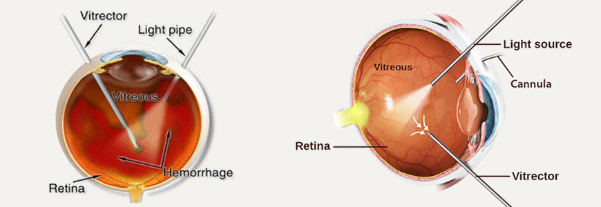
Any surgery done for treating a disease of the Vitreous or Retina involves removal of the Vitreous Gel and is called "Vitrectomy". The vitreous can be reached only by making cuts in the walls of the eye. This is a very delicate operation performed with an operating microscope and special needle - sized instruments. The most common indication for this operation is removal of the Vitreous, which has lost its transparency and, therefore, has become an obstacle to the incoming light. In this surgery most of the non - transparent vitreous is removed and replaced with a clear solution. Vitrectomy may also be used to remove the pulling forces of the Vitreous, which may have led to detachment of the retina. This operation may also be used to Remove Blood Clots, Infectious Material, Cataract, Foreign Bodies, and Abnormal Membranes from the interior of the eyeball. Sometimes it is done for diagnostic purposes for diseases of unknown origin. Occasionally it may be necessary to Inject Air, Gas, or Silicone Oil into the eye after removing the Vitreous Gel.
How is the operation performed ?
The surgery may be done under general anaesthesia ( sound asleep ) or under local anaesthesia ( you are awake but feel no pain ). The operation takes two to four hours. Usually one operation is Sufficient, Occasionally Additional Surgery may be required. Most patients stay in hospital for one or two hours longer hospitalization may sometimes be necessary. A face - down position for sleeping may be suggested for several days. The operated eye will be bandaged for one day. Occasionally both eyes may need to be bandaged to ensure complete ocular rest.
What may I do after the operation ?
For the first two weeks you should rest at home. Travelling should be avoided except to see the doctor. If gas has been injected into the eye, you should avoid air travel for several weeks until specifically authorized by the doctor. Postoperative instructions will be given to you at the time of discharge and these should be strictly followed. Most patients are able to return to their routine in four weeks.
What are the chances of success ?
The vision improves to some degree in 90% of simple Vitrectomy cases. In difficult cases however, improvement is seen in approximately 60% of the cases while in others it may remain the same or even decrease. The final degree of clarity of vision is usually not evident for about three months. How much vision a patient will ultimately have is difficult to predict in individual cases. Patients are usually able to see large objects but fine vision and reading vision may not improve.
What are the side effects and possible complications of surgery ?
Blurred Vision, Pain in the Eye, Redness and Swelling, Mucus Discharge and Enlaged Pupil and Double Vision are usually the temporary side effects and they clear up in a few weeks. Usually there are no complications, but some patients may have problems such as Recurrent Bleeding, Infection, or Elevated Pressure in the eye. Rarely, a retinal detachment or cataract may develop requiring further surgery, either during or after the Vitrectomy Operation.
Problems with Conventional Vitrectomy : The standard approach, which has been followed for the last 30 years, involves making 3 cuts around 1mm in size each, in the coats of the eyeball. But these relatively large cuts must be closed at the end of the surgery by taking sutures. Besides, the outermost covering of the eyeball, called the "Conjunctiva", also needs to be cut and sutured in this approach. This excess trauma along with the sutures taken, cause lot of Redness, Watering and Discomfort to the patient in the post - operative period.
What is sutureless vitrectomy ?
Recently, a new method of performing Vitrectomy has been invented and is becoming popular all over the world. Similar to the developments in Cataract Surgery, Even Vitrectomy can be done with a "Sutureless" approach! This requires the use of special equipment that enables the surgeon to reach the vitreous using cuts that are as small as 0.5mm. These cuts close automatically and Do not require sutures.
Advantages of sutureless Vitrectomy : Since there are no sutures taken, there is no suture - related foreign body sensation or watering. Also, there is no need to cut the conjunctiva and so there is lesser redness of the eyes and faster recovery of the patient. All this increases the comfort of the patient in the post - operative period and he / she can resume his / her routine work at the earliest. Not only that, the overall time required for surgery is also less.
Where is it available ?
In India there are only a few places where such surgeries are being performed routinely. Our foundation is one of the leading centres of India and probably has the largest series of sutureless Vitrectomy cases since 2005.
Is it done for all patients ?
No. While the sutureless technique can be used for nearly all types of diseases, there are still a few conditions that will need the conventional Vitrectomy. After examining the patient, the retinal surgeon himself can decide whether it is possible to do the surgery with this special technique.